What is Sonography?

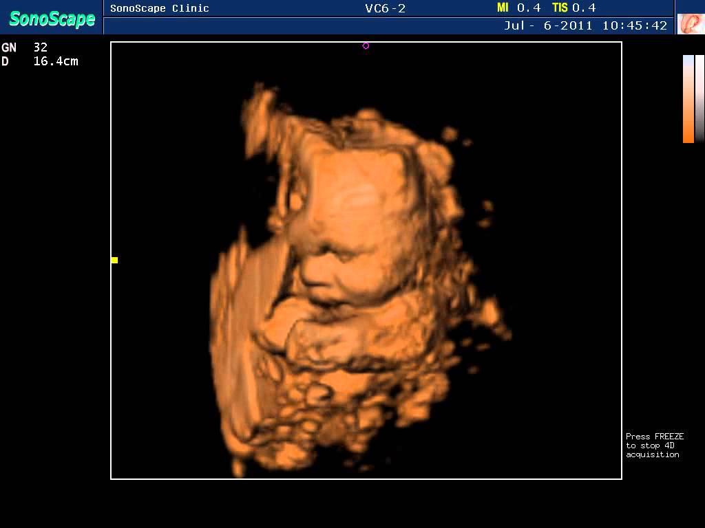
Sonography is a diagnostic medical procedure that uses high frequency sound waves to produce dynamic visual images of organs, tissues, or blood flow inside the body.
Sonography can be used to examine many parts of the body, such as the abdomen, breasts, female reproductive system, prostate, heart, and blood vessels. Sonography is increasingly being used in the detection and treatment of heart disease, heart attack, and vascular disease that can lead to stroke.

Quick Contact
Doctors use ultrasound in women, men, and children to gain advanced insights into the inner workings of the body. In fact, after x-ray exams, ultrasound is the most utilized form of diagnostic imaging available today.
Using ultrasound, doctors can monitor a variety of women’s health conditions from heart disease to breast abnormalities to several gynecological problems-accurately while limiting invasive procedures.
Ultrasound can help diagnose a wide variety of conditions in men, ranging from heart disease to abnormalities in the prostate gland or testicles. With children, doctors commonly use ultrasound to detect a variety of illnesses and disorders. A physician may use ultrasound to examine a child’s gastrointestinal tract for signs of appendicitis or a baby’s bone structure for alignment problems like congenital hip dislocation or spina bifida. An ultrasound exam of the head can detect hydrocephaly (water on the brain), intracranial hemorrahage (bleeding in the skull), and other conditions of the head.
Colour Doppler
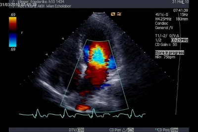
The advent of color Doppler was a breakthrough in medical ultrasound. With this technique it became possible to directly “observe” blood flow within the heart. Such visualization is achieved by color-encoding Doppler information and displaying the colors as an overlay on the 2D,3D and 4D image of the heart. The colors represent the speed and direction of blood flow within a certain area of the image (color box). The color box is divided into small sample regions (one color pixel). Each of these represent the mean velocity within the region as measured by multiple PW sample volumes. The mean velocity is then converted into a specific color. By definition, flow towards the transducer is depicted in red while flow away from the transducer is shown in blue. Different shades of red and blue are used to display velocity. Lighter shades of color are assigned to higher velocities. The exact manner in which velocities are displayed is defined by the so-called color map. Ultrasound scanners are usually provided with several maps from which the investigator may choose those he/she prefers. Some maps encode turbulent flow in yellow or green.
Our Services
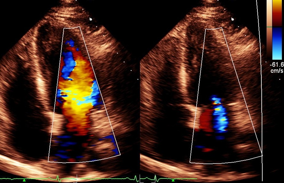
Turbulent flow is present when there are large variations in flow velocity within the sample region. The information is consistently updated from frame to frame, thus providing a clear view of how blood “flows”. The main advantage of color Doppler is the fact that one can view blood flow simultaneously in many regions of the heart. However, in contrast to PW and CW Doppler, color Doppler only permits semiquantitative assessment of blood flow velocities.